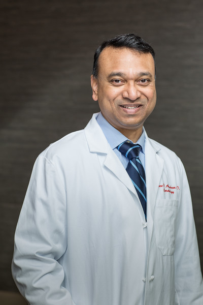
Faculty Spotlight: Theodore Abraham, MD

Hypertrophic cardiomyopathy (HCM) is the most common form of inherited heart disease, and too often is a silent killer – claiming the lives of young athletes and others who appear completely healthy. Echocardiographer Dr. Theodore (“Ted”) Abraham is on a mission to transform the way we diagnose and treat this condition.
Dr. Abraham was born in India, where both his parents were physician-scientists. He found cardiology appealing because it offered many ways to help patients, and subspecialized in echocardiography, which uses ultrasound images to study the heart. “I’ve always been a visual person, and echo involves a lot of images,” he said. “It also allows for good work-life balance, and allows me to do a fair amount of research.”
He earned his MBBS and medical degree from Goa Medical College at the University of Bombay, then came to the United States, where he completed his internal medicine residency and an echocardiography research fellowship at Wake Forest University in Winston-Salem, N.C. Dr. Abraham then completed his clinical cardiology fellowship at the University of Texas Southwestern, followed by an echocardiography and muscle physiology fellowship at the Mayo Clinic in Rochester, Minn.
The Mayo Clinic had a large clinic devoted to HCM. The disease causes the heart muscle to become abnormally thick – which can make it difficult for the heart to pump blood, and in some cases can lead to electrical problems that cause life-threatening abnormal heart rhythms, called arrhythmias. HCM often goes undiagnosed, because the disease may not cause noticeable symptoms before death.
“I started going to clinic with some of the world leaders in HCM, and became intrigued by the condition,” said Dr. Abraham. “Imaging plays a huge role in diagnosing, monitoring, and identifying the best therapy for the disease, and if a patient has surgery, determining whether the procedure was successful or not.”
Dawn of a New Imaging Tool
One obstacle to assessing heart health were the limited ways to measure muscle function. Until recently, the only two methods were to either cut out a piece of the heart and study it in the lab, or to sew metal beads onto the heart during open-heart surgery and use X-ray imaging to measure how fast the beads moved. These methods were far too invasive to be used routinely.
But in the early 2000s, as Dr. Abraham started his training at the Mayo Clinic, a new technology called strain echocardiography was just developing. “Strain” is an engineering term that measures how stretchy something is – for example, if a rubber band stretches from one foot to one-and-a-half feet, it demonstrates a strain of 50 percent. While traditional echocardiograms depend on cardiologists to visually estimate how well the heart or valves function, strain echocardiography uses computational processing to study the heart’s movement in minute detail.
An echocardiogram uses sound waves that bounce off the heart and “echo” back to form pictures, a bit like the way a bat uses echolocation to navigate. Strain echocardiography uses advanced computing systems to identify unique acoustic signatures called “speckles” that are associated with a specific muscle location. The computer recognizes each speckle, and tracks these over time. When a muscle contracts, speckles on each end of the muscle come closer together, and when the muscle relaxes, the speckles move further apart. The computer has the ability to track thousands of speckles at once. Instead of a human estimate of how fast a muscle is moving, the computer can provide a specific measurement.
At the Mayo Clinic, Dr. Abraham spent two years in an anesthesia lab that was one of the first in the world to validate and apply strain echocardiography to examine muscle physiology. “In the cubicle right next to me was a visiting PhD from Norway who had written the computer algorithm for the strain echocardiography software,” he said. “I learned directly from the person who wrote the program! It became a very exciting project. If we had trouble, he’d say, ‘Okay, I’ll fix it.’”
Judging by Function, Not Appearance
Not only can strain echocardiography identify how well a single muscle moves – it can pinpoint subtle problems that conventional echocardiography completely misses. As a faculty member at Johns Hopkins University for 14 years, Dr. Abraham’s lab pioneered applications of this imaging technique to help transform the understanding of HCM.
For example, a 38-year-old patient with no history of heart disease came to the emergency room. In the cardiac catheterization lab, the team found that one of his cardiac arteries was 95 percent blocked. “The conventional [echo] method never showed anything, but we did this [strain echo] map and it looked abnormal,” said Dr. Abraham. “Clearly, a computer that can read 200 or 300 frames per second is better than the human eye, which reads about 15 frames per second.”
At Johns Hopkins, Dr. Abraham’s team also lead a number of dyssynchrony studies – identifying whether part of the heart is lagging behind the rest of the muscle. A healthy heart is like a rowing team, with the entire heart pumping together in a coordinated manner. “With disease, some people aren’t rowing as hard, and some people are rowing backwards while others are rowing forwards,” he said. With strain echocardiography, he can tell whether one part of the heart is just weaker than the rest, or if a segment is actually moving in a different direction – a more serious abnormality. If any part of the heart lags more than 70 milliseconds behind the rest, it causes problems. Such patients may be candidates for a biventricular pacemaker, which can help patients feel better and live longer.
Strain echocardiography also is important for monitoring patients’ heart function over time. “It can catch small perturbations that we previously identified only when it was too late,” said Dr. Abraham. “Before, patients with HCM and other forms of heart disease would come in, get an echo, and often be told, ‘You have nothing wrong – I’ve looked at your echo and it looks great,’” he said. “They’d come back 10 years later and be in really bad shape.”
The ability to measure small changes over time is important for managing many types of heart disease, not just HCM. For example, some types of chemotherapy can potentially damage the heart. “Oncologists want high sensitivity imaging,” said Dr. Abraham. “They need a number they can follow, not me saying, ‘I think it’s 40 or 50 percent.’ Maybe the first time it’s 49 percent, and the second time it’s 41 percent – that’s a big difference.”
Interestingly, about half of the people with heart failure have hearts that contract normally – but their hearts have difficulty relaxing in between beats. Strain echocardiography can also be used to diagnose and monitor this condition, called diastolic heart failure, which is responsible for a large portion of patients who are readmitted to the hospital. Hypertension is the main cause of diastolic heart failure, but unfortunately there are no other therapies for treating the condition other than controlling high blood pressure.
Patients with HCM also have very high pressures in their hearts, a condition that is treated by performing open-heart surgery to remove the extra heart muscle causing obstruction in the heart. A company in South San Francisco is planning a clinical trial for a new drug that could enhance the heart’s ability to relax, which could offer a medical alternative to such an invasive procedure.
“All of cardiology will move into functional assessment,” said Dr. Abraham. “Echocardiographers are going to go from morphologists into physiologists.” That means that how a heart works will be more important than what it looks like. He compares it to assessing the soundness of a building. “What you see on the surface is not the full story,” he said. “It may look fine on the outside, so I’ll give it one point for that, but now I’m going to see if the windows are insulated, and test whether the elevators, heating, air conditioning and plumbing are working.”
Creating a Precision Map of HCM
While at Johns Hopkins University, Dr. Abraham founded the Johns Hopkins Hypertrophic Cardiomyopathy Center of Excellence and Athletic Heart Clinic, and built it into one of the top centers in the country. His group conducted research from experimental models to small clinical trials, served as the core lab for multicenter studies, and published 30 peer-reviewed papers.
Dr. Abraham collaborates closely with his wife, Dr. Maria (“Roselle”) Abraham, who is also an HCM physician-scientist who focuses most of her energy in the lab. She directs and he co-directs the new UCSF HCM Center of Excellence.
“We hit a plateau at Hopkins, and we’re very passionate about doing something that will be a game-changer in HCM,” said Dr. Abraham. “UCSF has this combustible mix of people, equipment, technology and deep science that will allow us to make the progress we need. By partnering with electrophysiologists, stem cell scientists, molecular biologists, developmental biologists, radiologists and others at the CVRI, Gladstone Institutes, and all the other scientific experts within the UCSF family, we believe we will truly make a difference. Our goal is to see whether we can really revolutionize the understanding and treatment of HCM.”
Dr. Abraham is excited to combine the power of strain echocardiography with other tools, such as metabolic imaging – another type of functional imaging that can reveal the series of chemical reactions at the cellular level. “We will marry those tools with morphology, genetics and biomarkers, creating multiple points of input to create a precision map of HCM,” he said.
“Although genotyping is important, it’s not the full story for HCM,” said Dr. Abraham. “For half of the people with severe HCM, we haven’t found the causative gene. And just because the gene is present doesn’t mean you have the disease…. Even though HCM is thought to be a uniform condition, it’s really not. There are multiple types of HCM, and if we know what the fundamental pathway is for a specific individual, we might be able to stop you from developing derangements and symptoms. This is one of the first major changes in how HCM is understood.”
“After only the first six months of his tenure as chief of the echocardiography laboratory, it is not an exaggeration to say that Dr. Abraham has initiated changes in our clinical and research missions that put us squarely on track to be the leading laboratory in the country,” said Dr. Nelson B. Schiller, John J. Sampson-Lucie Stern Endowed Chair in Cardiology. “In addition to strong clinical leadership, Ted is nationally recognized in the care and federally funded investigation of HCM and as a leader in national medical organizations. Speaking as the founder of the UCSF Echocardiography Laboratory, I am thrilled with the accelerated evolution toward greater excellence that Dr. Abraham will ensure.”
In addition to his research and care of patients, Dr. Abraham serves as clinical chief of cardiology for inpatient operations. “I love clinical integration, and did a lot of that at Johns Hopkins,” he said. “I am excited to look at our operations, whether it’s the diagnostic labs or cardiology critical care, to make them more cohesive, efficient, forward-looking and to establish programs of distinction.”
In his spare time, Dr. Abraham enjoys biking with his daughter, traveling and exploring new restaurants.
“I’m really excited to be at UCSF,” he said. “So far, it’s been so heartening to interact with our team in echo and everyone else at the institution. People are out to be excellent and deliver quality. That’s really the culture of UCSF, and I look forward to partnering with everyone to take UCSF Cardiology to the next level.”
– Elizabeth Chur
