
Faculty Spotlight: Elyse Foster, MD
Dr. Elyse Foster, a pioneer in adult congenital heart disease and the field of echocardiography, has witnessed many transformations in the care of patients born with heart defects – as well as the field of medicine itself.
"The year I started medical school, 20 percent of the class were women," said Dr. Foster, Araxe Vilensky Endowed Chair in Medicine, who earned her medical degree at Tufts University School of Medicine in Boston in 1977. Just two years before, only 10 percent of the entering class were female. "With the women's movement, it was inspiring to see that [medicine] was something that many women were doing."
Dr. Foster was born in Brooklyn and grew up on Long Island. She attended Tufts as an undergraduate, originally planning to study early childhood education or special education. That all changed when she befriended a number of pre-med students who were women. By her sophomore year, Dr. Foster decided to pursue medicine, and majored in psychology. Through her volunteer work at a women's clinic in Boston and the emergency department at Massachusetts General Hospital, she also met women doctors for the first time.
She decided to pursue a career in cardiology because she enjoyed reading electrocardiograms and caring for heart attack patients, and had a number of inspiring cardiology mentors during residency, including Dr. Carl Apstein and Dr. Laura Wexler. Dr. Foster completed her internal medicine residency and cardiology fellowship at Boston University, where she was only the third woman to become a cardiology fellow. This disparity was reflected throughout the field. "We would go to cardiology meetings, and it would be seas of men," said Dr. Foster. "It was the only time you never had to wait in line for the women's room, because there were so few of us!"
Adult Congenital Heart Disease: Lifelong Care
She joined the Boston University cardiology faculty, then was recruited to the UCSF cardiology faculty in 1989. For many years, she directed the adult echocardiography laboratory – which uses ultrasound of the heart to diagnose and help guide treatment of patients at UCSF Medical Center. She also led the Adult Congenital Heart Disease Service for almost two decades, which cares for the burgeoning population of patients with heart defects who are now living into adulthood, thanks to improved surgical treatments.
"She is a giant in the field of echocardiography," said Dr. Jeffrey Olgin, chief of the Division of Cardiology and Ernest Gallo-Kanu Chatterjee Distinguished Professor in Clinical Cardiology. "In addition to building a world-class echo lab at UCSF and helping start one of the first adult congenital heart disease clinics, she has trained generations of echocardiographers."
"The whole field of adult congenital heart disease has grown from a very small field to an established subspecialty of cardiology," said Dr. Foster. "A lot of the operations for congenital heart disease were developed in the late 1950s and early 1960s, and there are still ongoing modifications to many of these surgeries. We've seen these patients grow up and create their own new natural history, because we didn't know what the long-term outcomes would be."
For example, in the early 1990s, cyanotic or "blue" babies whose heart malformations prevented their blood from being fully oxygenated had 90 percent mortality by age 10. Today, 90 percent survive until age 20. "What we couldn't anticipate were the late problems associated with their surgeries," said Dr. Foster. "For patients who were born with a single ventricle, would that ventricle fail? Was the [original] operation they had going to create so many long-term problems that their outcomes would be poor in the long run?"
Dr. Foster has followed a cohort of patients for 20 years, some of whom had heart surgery at UCSF, and others who have moved here as adults. Many of her patients are now in their 50s, and one of her oldest patients is now more than 70 years old. "It's an incredible field," said Dr. Foster. "Patients usually transition from their pediatric cardiologist to us between ages 20 and 25. We have the privilege of following many patients through marriage, pregnancy, childbirth, and hopefully into old age. It's a lifelong relationship."
Dr. Foster works with a large network of colleagues to care for these complex patients, including specialists in cardiac arrhythmias, electrophysiology and heart failure, as well as obstetrics and gynecology, pulmonology, and other fields. "This group of patients requires a village of physicians and health care professionals to care for them," she said.
Many patients with congenital heart disease have surgery as infants or children and enjoy relatively typical childhoods, but do not get adequate care once they leave for college or move away from home. "Because of the success of the procedures, they feel healthy, but are lost to follow-up until they get into trouble because suddenly something like an abnormal heart rhythm occurs," said Dr. Foster. "Then they present at some community hospital emergency room, and the people there don't understand their disease. Identifying these patients and getting them back into specialized care is really important."
Dr. Foster has been a leader in these efforts nationally. She co-wrote guidelines published by the American Heart Association (AHA) on how to best manage medical and psychosocial transitions to adulthood for adolescents with congenital heart disease. She also co-authored a scientific statement for the AHA on the diagnosis and treatment of older adults with congenital heart disease. In addition, she has served on the board of directors and medical advisory board of the Adult Congenital Heart Association, a patient advocacy group.
She has seen giant leaps forward in the care of adult congenital heart disease patients. Many patients have scar tissue on their hearts from prior heart surgeries, which can cause abnormal heart rhythms. Often, these can now be treated through ablation, in which an electrophysiologist strategically burns or freezes small areas of the heart to isolate rogue electrical currents. Another major innovation has been the development of catheter-based therapies to patch holes in the heart or replace malfunctioning heart valves. Because this approach is done by inserting devices into blood vessels and snaking them up to the heart rather than through open-heart surgery, they broaden the treatment options for patients who have already endured multiple open-heart surgeries as children.
Dr. Foster has been a lead investigator of the MitraClip system, which uses a device that is inserted through a catheter to repair a leaky mitral valve – a bit like using a tiny clothespin to secure loose valve flaps, preventing backflow of blood in the heart. Her group has also studied the effectiveness of biventricular pacing to treat heart failure, in which a special kind of pacemaker helps the heart contract in a more coordinated and efficient way.
Solving Puzzles with Echocardiography
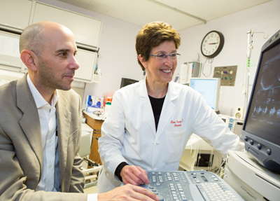
Dr. Foster is also an expert in echocardiography, which is used to diagnose and treat many forms of heart disease. "Ultrasound of the heart is important for every aspect of cardiology," she said. "It provides a wealth of information about cardiac structure and function, and the pressure in the heart. It's also very useful, because it's non-invasive, relatively inexpensive, widely available, and portable – it can be done at the bedside, in the operating room or in the emergency room."
For example, "broken heart syndrome" or stress-induced cardiomyopathy was just identified a few decades ago. It more frequently affects women than men, and is characterized by chest pain and shortness of breath that is brought on by an emotionally or physically stressful event. Patients often think they are having a heart attack. However, the condition causes a signature ballooning in the bottom tip of the heart, which can be seen on an echocardiogram. "I had this patient in her 90s whose house had a rat infestation, and she was very upset about it and developed chest pain," said Dr. Foster. "I saw her in the emergency room, put the echo probe on her chest, and said, ‘I know what you have!' I have seen her a couple times since, and she has perfect heart function now."
When teaching trainees, Dr. Foster emphasizes the importance of using echocardiography and other tools to better understand the big picture. "More than anything, we need to place the results of the echocardiogram in the context of the patient and their clinical problem," she said. "What is the chief complaint, what do we know about underlying diseases, and how can we integrate the findings on the echocardiogram into the diagnosis and care of the patient? When things don't fit together, you've got to work out that puzzle."
For example, she and her colleagues published a case in the New England Journal of Medicine about a patient with severe heart failure of mysterious origin. The echocardiogram showed that the heart was actually squeezing very well, which didn't make sense given her other symptoms. They eventually discovered that she had an arteriovenous fistula – a rare condition in which the arteries and veins are directly connected, rather than being separated by small blood vessels called capillaries. A UCSF vascular surgeon performed a nine-hour surgery to repair the fistula, and the patient's heart failure melted away. "It was important to look at the echocardiogram with a very critical eye and say, ‘Wait a second, something is out of the ordinary,'" said Dr. Foster. "It's an example of putting all the data together and paying attention to subtle findings."
Dr. Foster is an enthusiastic teacher, and has mentored generations of cardiologists. "Sitting down with an attending like Dr. Foster and having her go through each echo from start to finish is amazing," said Dr. Nalini Colaco, a first-year cardiology fellow at UCSF. "There were so many things that I wouldn't be able to pick up on my own that she showed us how to work through. And when she sees an interesting echo, she'll call us all over and say, ‘Look at this! It's something you should see and know.' You can really sense when someone wants to teach you, and when they are so willing to give their time to engage with you."
"It has been a privilege to train and work with Dr. Foster," said Dr. Ian Harris, director of the UCSF Adult Congenital Cardiology Program. "In addition to being a world-class echocardiographer and top-notch clinician, she is an exceptional teacher and mentor. I owe my entry into the field of adult congenital cardiology to her, and am indebted to her for her invaluable support and guidance over the years."
"During nearly two decades as chief of the UCSF Echocardiography Laboratory, Elyse Foster has melded extraordinary administrative instincts and exceptional clinical acumen," said Dr. Nelson B. Schiller, John J. Sampson and Lucie Stern Chair in Cardiology, who founded the UCSF Echocardiography Laboratory and the UCSF Adult Congenital Heart Disease Clinic. "As a result, the laboratory has maintained its position of world leadership while providing unwaveringly excellent services. The extraordinary growth in demand for echocardiography examinations of ever-increasing complexity stands as testimony to Dr. Foster's effective stewardship. In parallel, she has developed a competitive advanced training program for post-fellowship physicians seeking training in congenital and standard imaging. UCSF Cardiology has been most fortunate to have benefitted so richly from her dedicated efforts."
Looking to the Future
Dr. Foster plans to retire later this year, but will continue to work part-time in the echocardiography lab, see patients with congenital heart disease and structural heart defects, and continue her research into better treatments for valve disease. She also plans to continue her collaboration with the Adult Congenital Heart Association to promote education for physicians and adults with congenital heart disease and improve transitions between pediatric and adult cardiology for such patients. In addition, she will continue her work with the California Department of Public Health to reduce pregnancy-related mortality related to congenital heart disease.
Dr. Foster's entire family is involved in medicine: her husband, Dr. William Shalen, recently retired as a physician at Kaiser Permanente Medical Center in Richmond; her daughter, Dr. Julia Shalen, graduated from the UCSF School of Medicine and is currently a pediatric intern at UCSF; and her son, Dr. Evan Shalen, graduated from Boston University School of Medicine and is now a second-year internal medicine resident at UCSF. Dr. Foster is also excited to spend time with her first grandchild, Finn, who was born in 2015.
– Elizabeth Chur